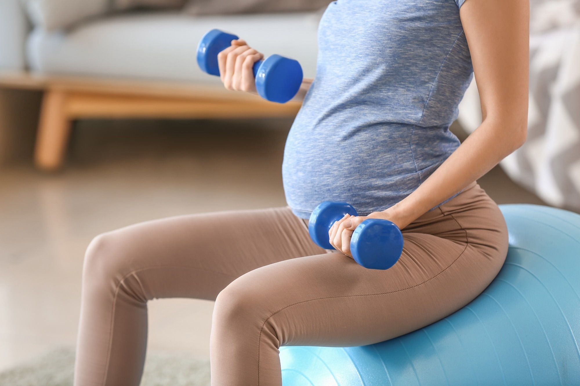
In a longitudinal study published in Scientific Reports, researchers investigate the effect of maternal physical activity on the size and growth of the yolk sac during early pregnancy. To this end, maternal physical activity was found to affect human yolk sac development, with this effect dependent on gestational age and embryonic sex.
 Study: Maternal physical activity affects yolk sac size and growth in early pregnancy, but girls and boys use different strategies. Image Credit: Pixel-Shot / Shutterstock.com
Background
The yolk sac is a structure observed during the early development of the human embryo that provides nutrients and allows gas exchange to the fetus until the placenta develops fully. Moreover, the yolk sac also plays a role in overall developmental processes such as protein synthesis, gastrointestinal tract formation, stem cell production, and hematopoiesis.
Various maternal factors such as height, weight gain, and sleep duration are known to affect yolk sac size. Maternal physical activity, for example, is a modifiable lifestyle factor associated with glucose control, weight gain, and overall outcomes for the mother and the fetus.
About the study
The present study aimed to understand how maternal physical activity potentially affects the intrauterine environment and early embryonic development, as indicated by the size and growth of the yolk sac.
As a part of the ongoing conception-implantation interval in pregnancy (CONIMPREG) research program, this prospective and longitudinal study included 196 healthy and nonsmoking women with regular menstrual cycles who could conceive naturally. The women were between 20 and 35 years old, and their body mass index (BMI) was between 18 and 30 kg/m2.
The assessment of participants was conducted at four study visits. During the first visit, prior to conception, height and body composition were measured, and physical activity was recorded using an actigraph, a wireless and noninvasive monitor.
In the second visit at seven weeks of gestation, embryo viability, gestation length, and yolk sac were assessed through ultrasound imaging. At 10 weeks, yolk sac measures were repeated. During the final visit at 13 weeks, maternal physical activity and body composition were reevaluated.
The statistical analysis involved using ordinary least square linear (OLS) regression models, analysis of variance (ANOVA), and the estimation of means, standard deviations, minima, and maxima.
Study findings
The mean gestational length was 281 days according to the last menstrual period (LMP) date or 278.5 days, as reflected by the crown-rump length (CRL) of the embryo in the first trimester. The pregnancies showed a lower rate of complications, including preterm birth, gestational diabetes, a five-min Apgar score of seven or less, and gestational hypertension at 3.2%, 3.7%, 1.1%, and 3.2%, respectively.
The total activity duration was five hours and 55 minutes prior to conception, which was reduced by one hour and 36 minutes by the end of the first trimester. This pattern was consistent between light and moderate-vigorous activity.
The mean yolk sac size was found to be 4.7 mm at week seven, which significantly increased to 5.9 mm at week 10 with individual variability. The reproducibility of the ultrasound measurements of the yolk sac size was assessed by measuring the intra- and interobserver variabilities, which were 0.08% and 0.09%, respectively, thus confirming the measurement precision.
When gestational and embryonic age were not considered, the yolk sac size was not significantly affected by maternal physical activity. However, at week seven of gestation, an increased preconception physical activity was associated with a larger yolk sac diameter in male embryos, with no such effect on female embryos.
At week 10, both embryonic sexes were affected by maternal physical activity. While male embryos showed a negative association between yolk sac size and maternal physical activity at this stage, female embryos showed a strong positive correlation in the two variables, with a 24% larger yolk sac than male embryos.
A significant interaction was also observed between embryonic sex and the daily physical activity of the mother. The effects at 13 weeks of gestation were not statistically significant. Additionally, preconception maternal physical activity was associated with variations in yolk sac growth velocity, with a significant difference observed among the two embryonic sexes.
The study’s findings are strengthened by its prospective design, the inclusion of preconception data of healthy women with natural and low-risk pregnancies, and the statistical models used for analysis. Notable limitations of the study include its relatively low generalizability, as well as the lack of continuous measurement of physical activity and control of the impact of maternal nutrition and stress.
Conclusions
The findings reiterate that maternal cues may influence human embryonic growth and development. Maternal physical activity was found to affect yolk sac size in a graded and embryonic sex-specific manner.
During early pregnancy, this effect was found to be in short phases. Further research is warranted in this area to understand the impact of maternal physical activity on the offspring’s long-term health.
Journal reference:
- Vietheer, A., Kiserud, T., Ebbing, C., et al. (2023). Maternal physical activity affects yolk sac size and growth in early pregnancy, but girls and boys use different strategies. Scientific Reports 13. doi:10.1038/s41598-023-47536-4, ]

 PARENTING TIPS
 PREGNANCY
 BABY CARE
 TODDLERS
 TEENS
TEENS HEALTH CARE
 ACTIVITIES & CRAFTS

