An article published in the journal PNAS describes the ecological causes of gut dysbiosis and its effects on human disease.
 Study: Gut dysbiosis: Ecological causes and causative effects on human disease. Image Credit: Kateryna Kon / Shutterstock
Background
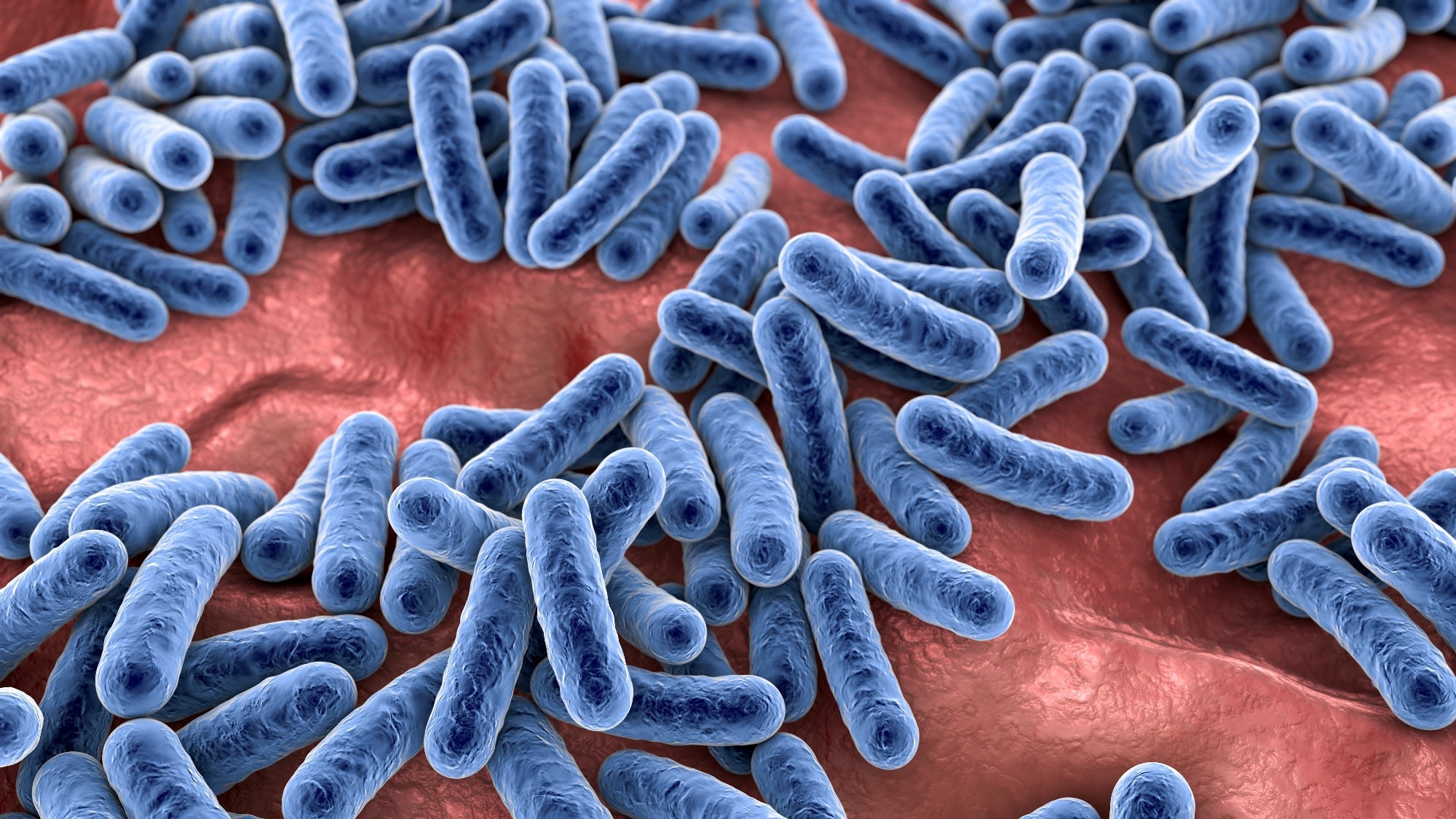
The gut microbiota refers to the vast number of microbial communities in the human colon. Secondary metabolites produced by these microbial communities play vital roles in health and disease.
Gut dysbiosis refers to an imbalance in gut microbiota composition, which is known to be associated with various chronic diseases, including cardiovascular disease, diabetes, chronic kidney disease, inflammatory bowel disease, and colorectal cancer.
In this article, the authors have provided a detailed overview of the ecological causes of gut dysbiosis. They have also thoroughly analyzed the causative link between gut dysbiosis and human disease.
Association between host physiology and gut microbiota dysbiosis
High-throughput genomic screening is the most widely used method to study gut microbial composition and diversity. However, this method is not adequately suitable for fully understanding the causative factors associated with gut dysbiosis as it only addresses the gut microbes and their genes but does not analyze the impact of the host environment.
In normal physiological conditions, the gut microbiota is dominated by beneficial bacterial communities (Clostridia and Bacteroidia). However, infections induced by enteric pathogens, including Salmonella Typhimurium, Citrobacter rodentium, and Toxoplasma gondii, can cause intestinal inflammation, which subsequently can increase the abundance of pathogenic bacterial communities (Gammaproteobacteria and Bacilli).
Breakthrough studies investigating the mechanism of colitis-induced growth of Salmonella Typhimurium have found that phagocytes recruited into the intestinal lumen during gut inflammation provide respiratory electron acceptors for the pathogen by oxidizing endogenous sulfur compounds to tetrathionate.
A collective analysis of bacterial and host genetics has made it possible to understand how host-derived tetrathionate during colitis can facilitate pathogen growth within the gut microbiota.
Nitrate was also identified as a host-derived respiratory electron acceptor during pathogen-induced colitis. An increased nitrate concentration in the gut during phagocyte recruitment was found to facilitate anaerobic respiration of commensal bacteria, including Salmonella Typhimurium and Escherichia coli.
Pathogen-induced colitis increases the diffusion of host-derived oxygen into the gut, leading to a loss of anaerobiosis. Antimicrobials released by luminal phagocytes reduce the microbial density and alter the microbiota composition by reducing the abundance of bacteria that produce short-chain fatty acids butyrate.
A reduction in butyrate concentration results in a reduction in mitochondrial oxygen consumption by gut epithelial cells and a shift of energy production toward aerobic glycolysis and, subsequently, induction of epithelial oxygenation. This increases oxygen flow from the epithelial surface, facilitating the pathogen growth in the gut through aerobic respiration.
Ulcerative colitis-induced gut dysbiosis is characterized by an increased abundance of Gammaproteobacteria and a reduced abundance of Clostridia. Available clinical data and animal experimentation data indicate that the increased abundance of pathogenic anaerobic bacteria in ulcerative colitis is caused by host-derived oxygen and nitrate. In other words, these observations indicate that the increased availability of host-derived respiratory electron acceptors is an ecological cause of gut dysbiosis in ulcerative colitis.
Regarding the association between antibiotic therapy and gut dysbiosis, evidence indicates that antibiotics-induced depletion in gut microbiota-derived short-chain fatty acids causes metabolic reprogramming in gut epithelial cells, which subsequently increases the availability of host-derived oxygen and nitrogen in the gut.
This increased availability of respiratory electron acceptors is associated with an increased abundance of pathogenic Escherichia coli during antibiotic therapy.
Association between gut microbiota dysbiosis and disease
The pathophysiology of many chronic diseases is associated with changes in gut microbiota composition and activity during dysbiosis.
In inflammatory bowel disease, microbiota dysbiosis is characterized by an increased abundance of pro-inflammatory Enterobacteriaceae. In mouse models of colitis, administration of sodium tungstate has been found to selectively reduce the expression of Enterobacteriaceae and suppress intestinal inflammation. These observations indicate that a high abundance of Enterobacteriaceae as a signature of dysbiosis is causatively associated with increased gut inflammation during inflammatory bowel disease.
Similarly, studies have found that dysbiosis triggers tumorigenesis of the gut microbiota by specifically increasing the abundance of colibactin-producing Escherichia coli. Colibactin is a genotoxin, and colibactin-producing Escherichia coli plays a direct role in triggering oncogenic mutations in patients with colorectal cancer.
Microbial metabolism during dysbiosis
Trimethylamine (TMA) is a harmful metabolite produced by the gut microbiota during the catabolism of red meat-derived nutrients choline and carnitine. TMA is absorbed and metabolized by the host to produce trimethylamine-N-oxide (TMAO).
TMAO is a uremic toxin, and patients with cardiovascular disease, chronic kidney disease, and type 2 diabetes exhibit high plasma levels of TMAO. Targeted inhibition of microbiota-induced TMA production has been found to attenuate atherosclerosis and chronic kidney disease in mice. These observations highlight the causative link between gut microbiota-derived metabolites and human morbidity and mortality.
Overall, this article describes that increased availability of host-derived respiratory electron acceptors can change the composition and function of gut microbiota by controlling microbial growth resources.

 PARENTING TIPS
 PREGNANCY
 BABY CARE
 TODDLERS
 TEENS
TEENS HEALTH CARE
 ACTIVITIES & CRAFTS

