In a recent study published in Scientific Reports, researchers identified shared genetic markers and immune system involvement in systemic juvenile rheumatoid arthritis (sJRA) and type 1 diabetes mellitus (T1D).
The study identified these by way of meta-analysis of microarray data analyzing the roles of transcription factors adenine and thymine (AT)-rich interaction domain 3A (ARID3A), Nuclear Factor, Erythroid 2 (NFE2), and Runt-related transcription factor 3 (RUNX3) in these diseases.
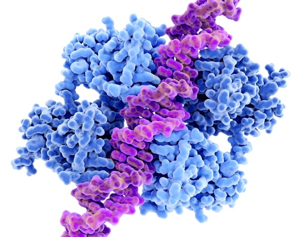
 Study: Exploration of common genomic signatures of systemic juvenile rheumatoid arthritis and type 1 diabetes. Image Credit: Khongtham/Shutterstock.com
Background
Juvenile idiopathic arthritis (JRA) is a leading chronic rheumatic disease in children, encompassing various diseases with diverse genetics, progressions, and outcomes.
Its classification is ever-evolving due to its heterogeneity. sJRA is especially concerning due to severe complications and varied early symptoms leading to diagnostic challenges.
T1D dominates pediatric diabetes cases, with rising incidence in younger children and increased complication risks, especially in China, where acute complications like diabetic ketoacidosis are alarmingly high.
By analyzing gene expressions in peripheral blood mononuclear cells (PBMC), research shows a genetic link between JRA and T1D, specifically highlighting the connection between sJRA and T1D.
Considering the health risks for children, it is essential to conduct further research on shared genetic indicators to enable early diagnosis and intervention for those highly susceptible to the coexistence of these diseases.
About the study
Gene expression profiles related to JRA and T1D were sourced from the Gene Expression Omnibus (GEO) database using the Medical Subject Headings (MeSH) “Arthritis, Juvenile Rheumatoid” and “Diabetes Mellitus Type 1.”
These profiles were filtered based on three criteria: they had to include sJRA, T1D, and control groups; originate from the PBMC tissue source; and contain analytical data.
A meta-analysis was then carried out on the datasets GSE7753, GSE21521, GSE193273, and GSE55100 using the R package “ExpressAnalystR.” The process involved individual dataset analysis applying the Benjamini-Hochberg’s False Discovery Rate (FDR) with p-values < 0.05.
After merging datasets post-annotation, the batch effect was adjusted to enable unbiased analysis. Fisher’s method identified Differentially Expressed Genes (DEGs) with a significant value of <0.05.
To construct the Transcription Factors (TFs)-target SDEGs network, 1665 TFs were downloaded from Human Transcription Factor Database (HumanTFDB). Using the hTFtarget database, potential target genes of these TFs within the SDEGs were pinpointed.
Only those with complete evidence were considered, and the network was visualized via Cytoscape (3.9.0).
Functional enrichment analysis of the gene sets was conducted using c, tapping into databases such as Kyoto Encyclopedia of Genes and Genomes (KEGG), Gene Ontology (GO), Reactome (REAC), and WikiPathways (WP).
Lastly, Cell-type Identification by Estimating Relative Subsets of RNA Transcripts (CIBERSORT), an R package, was employed to examine immune cell infiltration in sJRA and T1D samples. GSE7753 and GSE9006 datasets were specifically used for this analysis due to the specific data transformation requirements of CIBERSORT.
Study results
The data revealed the presence of 245 down-regulated SDEGs and 175 up-regulated SDEGs in sJRA and T1D. When examining the nature of these differentially expressed genes (DEGs), it was observed that the majority were associated with extracellular proteins.
Out of the complete set of SDEGs, only a small subset, 6 up-regulated and 9 down-regulated, were unrelated to extracellular proteins.
Using the HumanTFDB as a reference, a comparative analysis pinpointed 13 TFs in the up-regulated SDEGs, and a considerably higher count of 40 TFs in the down-regulated SDEGs. RUNX3 emerged as the transcription factor with the most potential target genes in the SDEGs.
Following RUNX3, ARID3A and NFE2 were noted to have the next highest counts of target SDEGs. Interestingly, while RUNX3 was downregulated, both ARID3A and NFE2 showed upregulation. Additionally, ARID3A and NFE2 demonstrated mutual regulation, with ARID3A also regulated by RUNX3.
Functional enrichment of these genes highlighted their involvement in various pathways and processes. The up-regulated SDEGs were associated with 238 GO terms, 62 REAC pathways, and 17 KEGG pathways.
These genes played crucial roles in cell cycle processes, innate immune response regulations, neutrophil degranulation, and several key signalling pathways, including Janus kinase/signal transducers and activators of transcription (JAK-STAT) and phosphoinositide 3-kinase (PI3K).
On the other hand, down-regulated SDEGs were associated with a smaller set of 154 GO terms, 36 REAC pathways, and 17 KEGG pathways. These were predominantly involved with the adaptive immune system, encompassing functions like T cell activation, differentiation of various T cell types, and cytokine functions.
They also touched upon innate immune system processes related to natural killer cell functions and specific signalling pathways.
Regarding the transcription factors targeting the SDEGs, the pathways they enriched in were largely consistent with those enriched by the up-and down-regulated SDEGs. One noteworthy observation was that neutrophil degranulation, and the cell cycle process were closely linked.
Likewise, terms associated with T cell activation, differentiation, cytokine functions, and natural killer cell activities were commonly enriched between the downregulated SDEGs and the targeted SDEGs.
The analysis extended to assessing the infiltration levels of 22 immune cells in T1D and vsJRA samples using the CIBERSORT method.
Both sJRA and T1D samples exhibited increased levels of neutrophils and naive Cluster of Differentiation 4 T (CD4 T) cells while showing decreased CD4 memory resting T cells.
When comparing monocyte infiltration levels between the diseases and control samples, it was evident that sJRA had a significantly elevated monocyte level.

 PARENTING TIPS
 PREGNANCY
 BABY CARE
 TODDLERS
 TEENS
TEENS HEALTH CARE
 ACTIVITIES & CRAFTS

