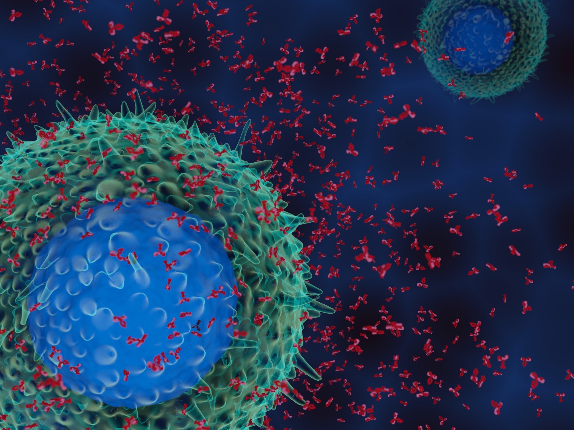
In a recent study posted to the medRxiv* preprint server, researchers examine a large dataset of patients with multisystem inflammatory syndrome in children (MIS-C) to determine whether their autoantibodies target a different set of host proteins as compared to healthier controls.
 Study: A distinct cross-reactive autoimmune response in multisystem inflammatory syndrome in children (MIS-C). Image Credit: Meletios Verras / Shutterstock.com

 *Important notice: medRxiv publishes preliminary scientific reports that are not peer-reviewed and, therefore, should not be regarded as conclusive, guide clinical practice/health-related behavior, or treated as established information.
Background
Severe acute respiratory syndrome coronavirus 2 (SARS-CoV-2) infection can lead to long-term dysregulation of the immune system and a broad spectrum of secondary infections and diseases, including post-acute sequelae of coronavirus disease 2019 (COVID-19) in adults. In children, while the outcomes of COVID-19 are generally mild, rare cases progress to MIS-C.
MIS-C symptoms, such as systemic infection, prolonged fever, conjunctivitis, rash, and, in some cases, coronary artery aneurysms and myocarditis, are similar to those that are reported in severe cases of Kawasaki Disease. However, MIS-C after SARS-CoV-2 infection also presents as cardiac dysfunction, GI problems, hematological complications such as lymphopenia and thrombocytopenia, and various other muti-organ complications.
Autoimmunity and alterations in adaptive and innate immune responses in MIS-C have also been linked to distinct cytokine and inflammatory signatures, with cross-reactivity of autoantibodies also implicated in MIS-C.
About the study
In the present study, researchers used cohorts of children with a history of COVID-19 with and without MIS-C to profile the autoreactive antibodies and antibodies targeting SARS-CoV-2 using phage immunoprecipitation and sequencing (PhIP-Seq), which has been extensively used in various diseases to identify novel autoantigens. A human proteome-wide library consisting of 768,000 elements, which has previously been instrumental in defining biomarkers for various diseases and identifying novel autoimmune conditions, was also analyzed.
The MIS-C cohort comprised 199 cases, while the at-risk cohort comprised 45 individuals who previously had SARS-CoV-2 infection but did not present with MIS-C. All patients had nucleic acid amplification-confirmed SARS-CoV-2 infection, while MIS-C patients had additional serology tests.
Split luciferase binding assays were used for the deoxyribonucleic acid (DNA) coding of peptides of interest. Specific DNA expression plasmids were used for the radioligand binding assays.
Logistic regression machine learning was used to identify the presence of differentially enriched peptides that distinguish MIS-C samples from at-risk control samples. The Kolmogorov-Smirnov test was used to identify statistically enriched autoreactivity. The validity of the findings was confirmed using an independent cohort comprising MIS-C patients and children severely affected by acute SARS-CoV-2 infection.
The results of PhIP-Seq were normalized to healthy controls to determine whether specific peptides were differentially enriched by the at-risk control or MIS-C samples. Furthermore, based on the prediction of preferential peptide display in the human leukocyte antigen (HLA) types associated with MIS-C, the researchers evaluated the presence of MIS-C-specific cross-reactive T-cells using peripheral blood mononuclear cells (PBMCs) isolated from MIS-C patients and at-risk controls.
Single-cell sequences from PBMCs obtained from individuals with asymptomatic, mild, or severe SARS-CoV-2 infections or influenza, as well as healthy controls, were used to analyze the T- and B-cell autoimmunity to SNX8, which is a key regulator of the MIS-C pathogenesis antiviral pathway.
Study findings
The antibody response in MIS-C patients was differentially reactive to SNX8 and a specific domain of the nucleocapsid protein of SARS-CoV-2 as compared to the antibody response of the at-risk controls. Furthermore, the SNX8 protein and this viral nucleocapsid region had remarkable biochemical similarities.
MIS-C patients with autoantibodies against SNX8 also had cross-reactive T-cells to the SNX8 protein and viral SARS-CoV-2 nucleocapsid region. These cross-reactive T cells are likely to contribute to immune dysregulation through the enlargement of SNX8-expressing immune cell lineages.
Fine epitope matching also identified the regular expression epitope [ML]Q[ML]PQG, which was similar in SNX8 and the viral nucleocapsid protein domain. This epitope also appeared to be shared by the B- and T-cells in MIS-C patients.
The SNX8 protein is functionally linked to the mitochondrial antiviral signaling (MAVS) pathway, and MIS-C is thought to be associated with an increased autoimmune response by SNX8 against tissues with high MAVS pathway expression. The researchers believe these results indicate similarities with other diseases, such as paraneoplastic autoimmune disease, where exposure to a novel antigen results in autoimmunity.
Conclusions
MIS-C patients exhibit immune responses against a distinct SARS-CoV-2 nucleocapsid protein domain, which is associated with cross-reactivity to the SNX8 protein.
The SNX8 protein and SARS-CoV-2 nucleocapsid protein domain were found to share an epitope. Notably, targeting this epitope by both B- and T-cells suggests the involvement of molecular mimicry, which needs to be explored further.

 *Important notice: medRxiv publishes preliminary scientific reports that are not peer-reviewed and, therefore, should not be regarded as conclusive, guide clinical practice/health-related behavior, or treated as established information.

 PARENTING TIPS
 PREGNANCY
 BABY CARE
 TODDLERS
 TEENS
TEENS HEALTH CARE
 ACTIVITIES & CRAFTS

