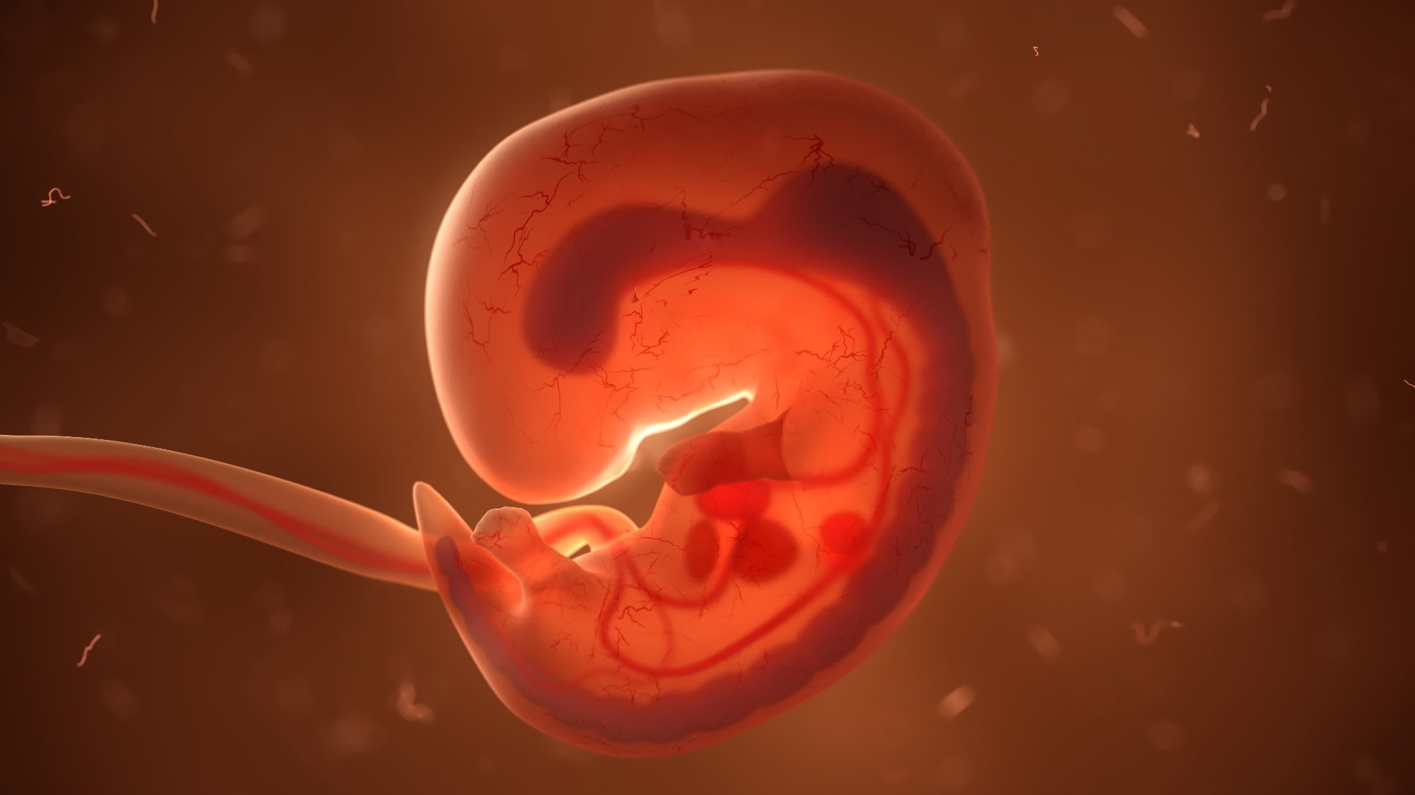
In a recent study published in the journal Science Immunology, researchers profile the origins and subsequent differentiation of embryonic and fetal immune cells during human lung development.
 Study: Early human lung immune cell development and its role in epithelial cell fate. Image Credit: u3d / Shutterstock.com
What do we currently know about fetal immune development, and why isn’t that enough?
Previous research has extensively documented the functions of immune cells in regeneration, maintaining homeostasis, particularly in the intestine and testis, and somatic tissue development. Studies have aimed to elucidate the structure, subtypes, and function of epithelial and mesenchymal cells; however, a gap exists in researchers’ understanding of the processes and functional roles of lung immune cells.
Immune cells are some of the most critical cells responsible for an infant’s survival from birth onwards. Given the defenses provided by lung-associated mucosal immune cells against airborne pathogens and inhaled toxins, the current dearth of literature on the subject is surprising. A possible explanation for this gap in the literature may be due to the complex nature of cell differentiation during embryonic development and the historical lack of techniques capable of safely tracing these differentiations throughout pregnancy.
A critical question that remains unanswered is whether immune cells might have functions over and above defense – could they modulate or otherwise influence the development of the tissues wherein they reside? Answering this and similar questions pertaining to human lung development at both cellular and molecular levels may result in the genesis of novel clinical interventions designed to repair and regenerate lungs, thereby affording millions or even hundreds of millions of patients an alternative to lung transplantation.
Previous research has characterized human lung developmental morphology and classified the process into five temporally overlapping stages. These consist of the embryonic stage between four and seven weeks post conception (pcw), the pseudoglandular stage between five and 17 pcw, the canalicular stage between 16 and 26 pcw, the saccular stage between 24 and 38 pcw, and the alveolar stage from 36 pcw to 21 years of age.
The first three stages, specifically between five and 22 pcw, represent the least understood period of lung development despite their collectively covering the entire evolution of epithelial stem cells into almost functional lungs.
About the study
The present study aimed to evaluate the temporal progression of the fetal immune system and elucidate its potential role in modulating embryonic lung development. Human fetal and embryonic samples were acquired from the Human Developmental Biology Resource (HDBR) Joint MRC/Wellcome Trust grant.
Pregnancies terminated between five and 22 pcw were used to obtain fresh lung tissue with written consent from donors. Karyotypic analysis was performed to ensure that included samples were free from genetic abnormalities and represented ‘typical’ human embryonic growth.
Immunohistochemistry (IHC) of lung tissues was used to validate immune cell types and quantity across the first three stages of embryonic lung development. IHC analysis further contributed to evaluating the locations of observed immune cells and variations therein from five to 22 pcw.
To improve the accuracy and reliability of immune cell quantification, three-dimensional (3D) quantification using confocal microscopy followed by Imaris software analyses was performed. Computed 3D images were compared to 2D images at every time point across the study duration.
Lung tissue digestion followed by flow cytometry and fluorescence-activated cell sorting (FACS) were used to validate IHC quantification estimates, elucidate relative proportions of CD3+, CD4+, CD8+, and regulatory T cells (Tregs) as a proportion of CD45+ populations for the same developmental stage, and sort CD45+ cells as a precursor to single-cell ribonucleic acid (RNA) sequencing (scRNA-sq).
Moreover, scRNA-sq served the dual purpose of validating flow cytometry results and the molecular characterization of immune cells across samples. Cellular indexing of transcriptomes and epitopes sequencing (CITE-seq) was additionally employed to improve the resolution of the results.
Human embryonic lung organoids were also used for functional characterization experiments, including cytokine treatments, dual Suppressor of Mothers Against Decapentaplegic (SMAD) transcription assays, and macrophage, dendritic cell (DC) culture, and cytokine arrays.
All obtained data were subject to statistical analyses consisting of one-way Analysis of Variance (ANOVA), unpaired two-tailed t-tests, residual maximum likelihood analysis (REML), and Tukey’s post hoc multiple-comparison test.
Study findings
The scRNA-seq, IHC, and functional organoid assays revealed that immune cell populations varied significantly across fetal developmental stages. Progenitor and innate immune cells, including myeloid, innate lymphoid (ILC), and natural killer (NK) cells, predominated in early developmental stages but were gradually replaced by T- and B-lymphocytes. CD45+ cells were nearly ubiquitous across developmental stages and in all lung-associated tissue regions; however, their relative quantity varied across time and location.
Molecular characterization of immune cells revealed 77,559 transcriptomic profiles, 61,757 of which are novel to science. Annotation of these profiles followed by clustering analyses revealed 59 clusters representative of all known immune cell categories. Analyses of the progenitors of these categories resulted in the discovery of unexpectedly high ILC- and early lymphoid progenitor (ELPs) densities.
Taken together, these results suggest that immune cells follow a biphasic pattern during fetal development and a spike in abundance during eight and 20 pcw. Quantitivate polymerase chain reaction (qPCR) evaluations suggest that the 20 pcw peak may be partially due to vascular maturation.
B-cell maturation in lungs was revealed for the first time using IHC and single-molecule fluorescence in situ hybridization (smFISH) assays. This added to a growing body of evidence that the bone marrow is not the sole source of mature B-cells, which is contrary to previous scientific beliefs.
Immunoglobulin (Ig) isotype expression in tandem with clonal expansion assays revealed that, during development, lung mesenchyme and epithelium support B-cell homeostasis through the secretion of modulatory chemokines, including CCL28.
The collective output of transcriptomic and cytokine assays revealed the complex interactions of multiple immune cell-secreted cytokines, which, in turn, were functionally validated to affect epithelial cell differentiation. These results suggest that immune cells serve a dual purpose of defense and lung development during fetal development, thereby confirming previous hypotheses.
Conclusions
In the present study, researchers combined cutting-edge transcriptomic analyses with IHC to elucidate immune cells’ structural and functional roles during fetal and embryonic development. Evaluations of fetal immune cells during the five to 33 pcw period revealed that complete B-cell maturation occurs in embryonic lungs, which contests the prevalent belief that the bone marrow is the sole source of mature B-cell populations.
Interleukin-1 beta (IL-1β) was found to be extensively produced by widely dispersed myeloid cells. IL-1β, in turn, was found to modulate and promote epithelial stem cell differentiation, highlighting the dual role of immune cells in both defense and lung epithelial development.
Together, these findings provide an immune atlas of developing human lungs and suggest a role for fetal immune cells in guiding development of the lung epithelium.”
Journal reference:
- Barnes, J. L., Yoshida, M., He, P., et al. (2023). Early human lung immune cell development and its role in epithelial cell fate. Science Immunology. doi:10.1126/sciimmunol.adf9988

 PARENTING TIPS
 PREGNANCY
 BABY CARE
 TODDLERS
 TEENS
TEENS HEALTH CARE
 ACTIVITIES & CRAFTS

